Artificial Intelligence for Chest X-Ray Transforming Patient Care
A field report about AI for Chest X-Ray and its impact in the UK

Radiology is a medical specialty that plays a critical role in diagnosing and monitoring a wide range of conditions by interpreting medical images such as X-rays, CT scans, and MRIs. Among these, Chest X-Rays hold a special place due to their pivotal role in diagnosing respiratory and cardiovascular diseases.
Radiologists are facing numerous challenges in efficiently managing an ever-increasing workload while maintaining high standards of care. This is evident in countries such as the UK, where in their announcement for funding of digital health, the NHS notes that “over 600,000 chest X-rays [are] performed each month in England” in order to combat lung cancer, “the leading cause of cancer death in the UK”1. As a hospital group that sees over 6,500 of chest X-rays a year, we can relate to the pressing needs that the UK is experiencing. Fortunately, the integration of artificial intelligence (AI) is poised to reshape the landscape of radiology, particularly in the realm of Chest X-Rays, and is enabling our hospital group to be well-positioned at the forefront of this wave of innovation.
Current Challenges
Over the past few years, I have encountered a number of major challenges in the radiology landscape:
- High Workload: Radiologists are burdened with heavy workloads, resulting in potential delays in image analysis and reporting. This situation can lead to delayed diagnoses and treatment plans, adversely affecting patient outcomes. This is necessarily true for the German radiology landscape but also for other countries such as England and their National Health Service (NHS).2
- Expertise Shortage: The demand for radiological services often outstrips the availability of skilled radiologists. The scarcity of specialized expertise can contribute to diagnostic errors and hinder the timely delivery of reports.3
- Data Overload: The exponential growth of medical imaging data poses challenges in managing and analyzing vast amounts of information. Manual analysis becomes time-consuming and error-prone.4
- Variance in Interpretations: Different radiologists might interpret the same image differently, leading to inconsistencies in diagnoses and treatment decisions.5
However, Artificial Intelligence, particularly machine learning, has demonstrated its potential to revolutionize the field of radiology, offering solutions to address the challenges outlined above:
Efficient Triage and Prioritization: AI can be used to triage chest X-rays, identifying urgent cases that require immediate attention. This ensures that critical cases are prioritized, improving patient care and reducing delays.
Assistance in Diagnoses: AI algorithms can be trained to detect patterns associated with various lung and cardiac conditions, assisting radiologists in making accurate diagnoses. This collaboration between human experts and AI can enhance the reliability of interpretations.
Workflow Optimization: AI tools can streamline radiology workflows by automating routine tasks like image preprocessing and labeling. This optimization allows radiologists to focus more on complex decision-making tasks.
Consistency in Interpretation: AI algorithms offer consistency in image interpretation, reducing the variability caused by human subjectivity. This can lead to standardized reporting and better patient care.
I also believe that artificial intelligence will provide us with valuable services in image acquisition. Specifically, AI will reduce the workload of us radiologists by taking care of more and more time-consuming routine tasks so that we can focus on reporting complex examinations and on our patients in need of treatment.
How can AI in CHEST X-RAY help to overcome the challenges
That's why we are one of the first hospitals in Southern Germany to use AI solutions routinely in our day-to-day practice. We have been inspired by thought-leading initiatives like the UK NHS which has been actively exploring AI solutions to address radiology challenges for many years. Initiatives like the "NHS AI Lab" aim to create an environment where the most successful AI technologies can be easily procured and used by hospitals.6 In the context of radiology, AI models are being developed to assist in identifying abnormalities in chest X-rays, such as pneumonia, tuberculosis, and lung nodules. These tools are not intended to replace radiologists but to work alongside them, enhancing their capabilities and efficiency.
At our hospitals, we use a variety of different AI products, amongst those solutions for fracture detection and Chest X-Ray reading. For the latter, I am currently working with different products in the area of Chest X-Ray, provided by our platform partner deepc. I am always amazed by the precision of how findings are detected by AI solutions.
In the following case a 43-year-old man presented with a tiny pneumothorax after a bicycle accident:

The pneumothorax was marked by the AI solution and confirmed on a CT scan of the shoulder, in the partially included upper thoracic aperture, which we also had in place. A very interesting case that shows how AI provides more security, especially to young colleagues in duty hours.
Another example that shows AI tools are very valuable decision support systems is the case of an elderly lady who was admitted to the emergency room of the Artemed Clinic Munich South after an unobserved fall in the nursing home:

On duty, the AI solution, marked a right apical pneumothorax. Overall, a complex case, as overlays are very present, primarily due to skin folds.
In fact, in the additional performed CT scan of the cervical spine, we spotted the upper thoracic aperture and the ventroapical pneumothorax gap on the right, measuring just under 1 cm. When it comes to treating patients without long waiting times, i.e. quickly but safely, AI is very valuable, especially in giving younger colleagues additional security with maximum stress reduction. These examples have shown how AI can help in critical situations. However, it is also a very prominent tool to diagnose lung cancer, one of the most frequent cancer-related causes of death in Europe.
Value of the Deployment via an AI Platform
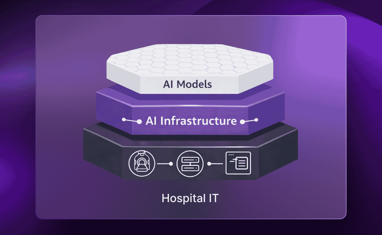
For the implementation of AI in our hospital there was only one choice - deepcOS, a globally leading Radiology AI platform. Using deepc's cutting-edge Radiology AI platform deepcOS, has been a game-changer in our hospital. The platform's scientifically curated vendor-neutral AI portfolio has empowered me to choose from a wide array of AI solutions that align perfectly with my diagnostic needs. What truly sets deepc apart is their seamless ability to swiftly roll out new AI solutions and seamlessly integrate them into my existing workflows and systems. It's remarkable how they provide not only the technology but also a unified framework for procurement, commercials, and thorough testing. With deepc, turning on a new solution feels like a breeze, and the value they add extends beyond just the technology – it's a comprehensive solution that enhances my efficiency, accuracy, and overall patient care.
AI's potential in radiology is vast, promising improved diagnostics and care. Amidst challenges like image overload and staffing shortages, AI, especially for chest X-rays, emerges as a solution. deepcOS simplified our journey, letting us test pre-curated AI solutions effortlessly, accelerating our selection process. deepcOS also enables streamlined AI integration, adherence to privacy protocols, and empowers quick algorithm selection. In this intricate field, deepcOS is a game-changer, simplifying AI adoption for a future of better patient care.
Interested in AI Chest solutions? Join our webinar to get a glimpse into the future of healthcare in the UK with AI, thanks to the NHS £21 million AI Diagnostic Fund here.
Reference
- (Department of Health and Social Care, 2023): https://www.gov.uk/government/news/21-million-to-roll-out-artificial-intelligence-across-the-nhs
- (RCR, 2023)
- (RCR, 2023)
- (More et al., 2023)
- (Benchoufi et al., 2020)
- (NHS, 2023)


.png)
.png)


.png)
